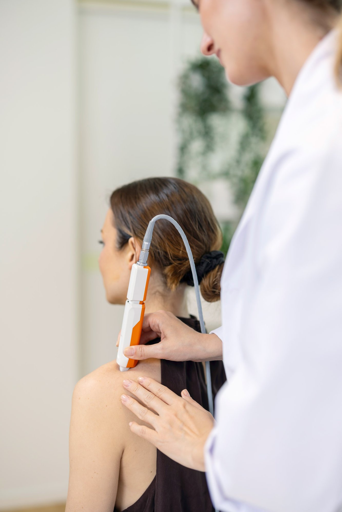
Early detection saves lives Melanoma is one of the most aggressive skin cancers – but also highly treatable when detected early. Nevisense supports healthcare professionals with objective, non-invasive diagnostic data to improve accuracy and confidence in clinical decision-making.
Rule out the risk of missing a melanoma with 97% sensitivity.
AI-powered diagnostic support
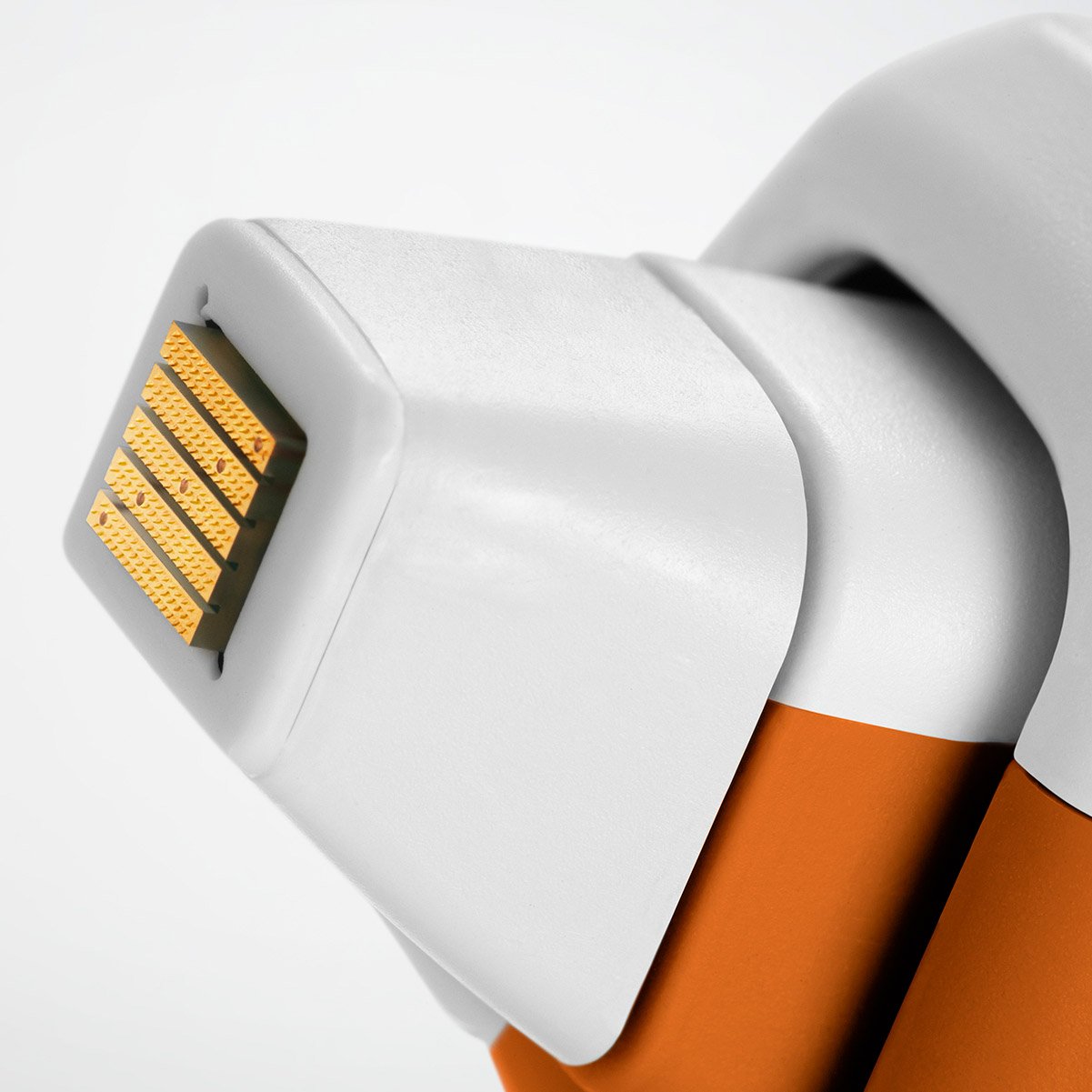
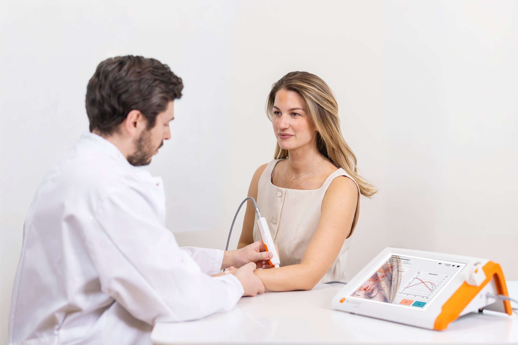
Nevisense combines electrical impedance spectroscopy (EIS) with AI to assess the skin’s bioelectrical signature, providing objective information beyond what the eye can see. The non-invasive assessment supports clinical decisions directly at the point of care. It provides a Nevisense score that helps clinicians evaluate suspicious lesions beyond visual inspection.
The Nevisense Melanoma score
The measurement provides 700 data points that are analyzed by the Nevisense proprietary algorithm (AI). Giving the clinician an actionable risk assessment score between 0-10. 0-3 indicate a negative score with a 99% negative predictive value for melanoma. 4-10 indicate a positive score with an increasing level of risk for melanoma. (Ref. 1)
Supporting dermatologists and skin cancer specialists
Nevisense is a valuable tool for dermatologists and clinics focusing on enhanced melanoma management and patient care.
Increase melanoma detection by 14% Additional insights into tissue properties help clinicians identify melanomas that may otherwise be missed (Ref. 2).
Identify melanoma with 97% sensitivity High sensitivity supports the detection of melanoma, including lesions with subtle visual features (Ref. 1).
Support ruling out melanoma with 99% NPV A high negative predictive value adds reassurance when assessing suspicious lesions, alongside clinical examination and dermoscopy. (Ref. 1)
Nevisense essentially offers two key advantages: First, fewer unnecessary excisions. Second, in my experience, a detection rate of nearly 100%.
Prof. Dr. Axel Hauschild
Dep. of Dermatology, UKSH, Kiel, Germany
Nevisense supports more informed biopsy decisions Studies show that the additional information provided by Nevisense helps physicians make more accurate biopsy decisions across all levels of experience. (Ref. 2)
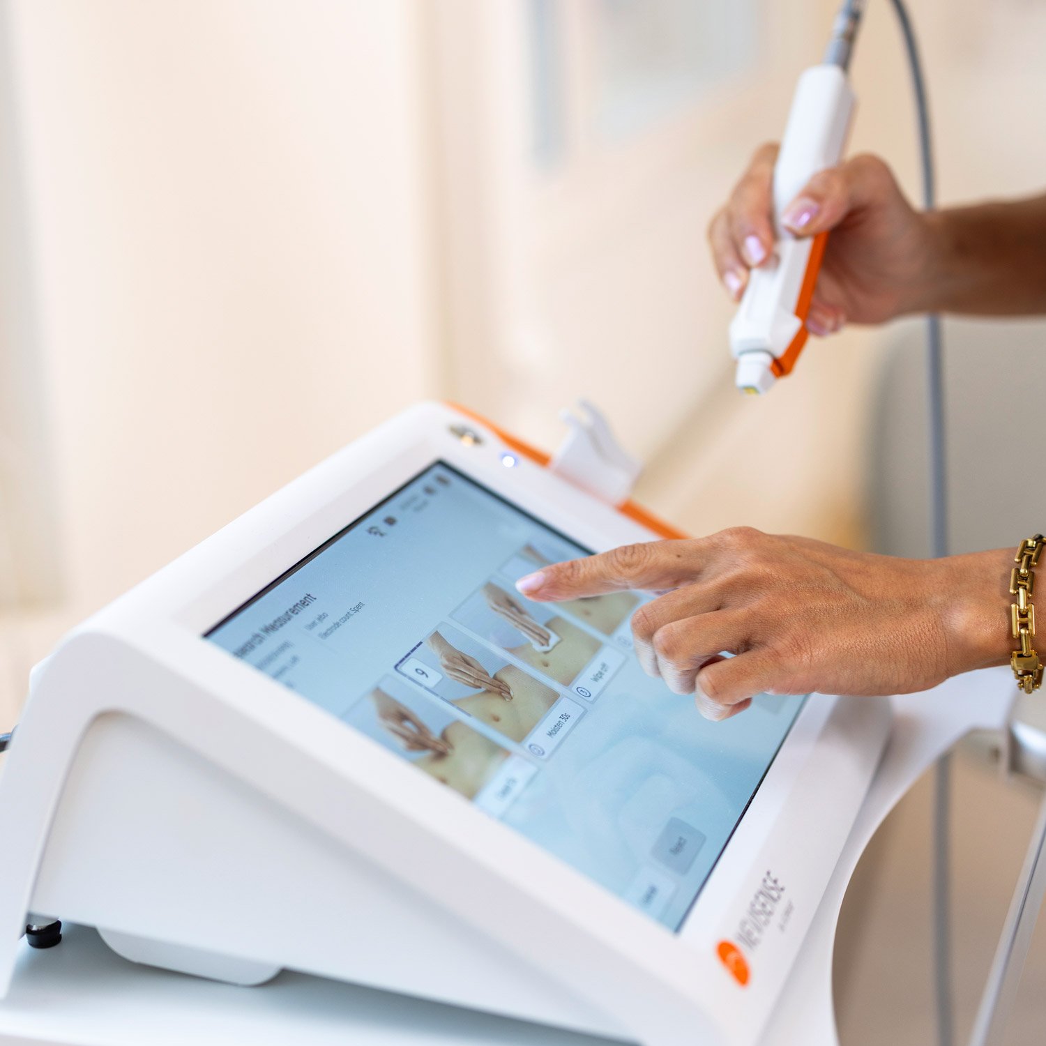
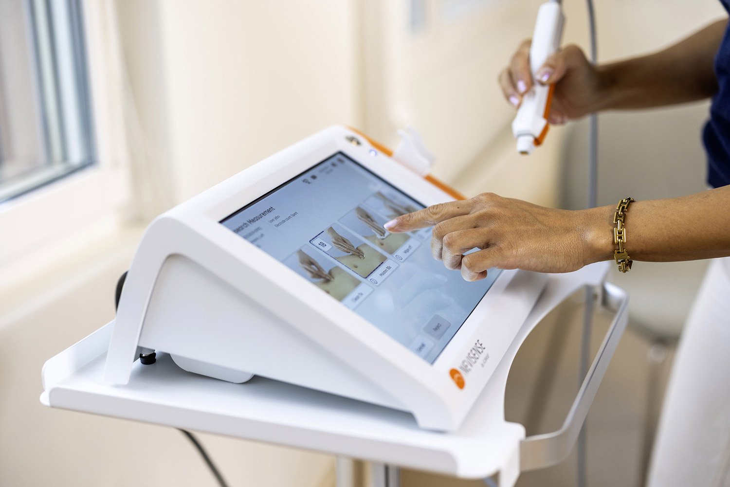
Seamless clinical integration The measurement process is non-invasive and easy to integrate into dermatology workflows, with results at the point of care for more well informed decisions.
Included in guidelines
Nevisense EIS technology is recognized in European and US clinical guidance as a complementary technology that can support the assessment of atypical skin lesions.
German S1 Guideline
The first German guideline on imaging diagnostics for skin diseases describes EIS as a potentially valuable clinical decision-support tool for assessing melanocytic and keratinocytic lesions. Read the guideline
EADO Melanoma Guideline
The European melanoma guideline reports a sensitivity of 97.7% and describes EIS as a potentially useful complementary tool in clinical practice. Read the guideline
US Expert Consensus
A US expert panel concluded that EIS can significantly enhance diagnostic assessment and clinical decision-making for clinically indeterminate pigmented lesions—including melanoma—with SORT Level A evidence. Read the consensus report
NCCN Guidelines
The NCCN Guidelines identify EIS among the diagnostic-support technologies that may aid in detecting new primary melanoma, particularly in patients with numerous or clinically atypical nevi. Visit NCCN
BOOK A DEMO
Discover how Nevisense can enhance your melanoma management
Book a personalized demonstration to learn how Nevisense can complement your current diagnostic process and provide objective support for clinical decision-making.
References: 1. Clinical performance of the Nevisense system in cutaneous melanoma detection: an international, multi-center, prospective and blinded clinical trial on efficacy and safety, Malvehy et al BJD, 2014 DOI: 10.1111/bjd.13121. 2. Integrating Electrical Impedance Spectroscopy into Clinical Decisions for Pigmented Skin Lesions Improves Diagnostic Accuracy: A Multitiered Study’, Litchman, Rigel et al, SKIN Vol4, Issue 4, July 2020

 Rule out melanoma with 99% NPV