
Här är det bara att börja fylla på innehåll.
Instruktion
Jag har lagt till sidorna även i huvudmenyn så det är lättare för dig att nå sidorna. Det tar vi bara bort sedan när det är klart.
Nedan innehåll är en klon på den generella references sidan som finns idag.
Rooted in academic research
Backed by clinical evidence
Peer-reviewed publications support Nevisense's clinical performance in assessing melanoma and keratinocyte cancers. Studies conducted across Europe, the US, and Asia demonstrate its value across a range of clinical settings.
Improving diagnostic accuracy
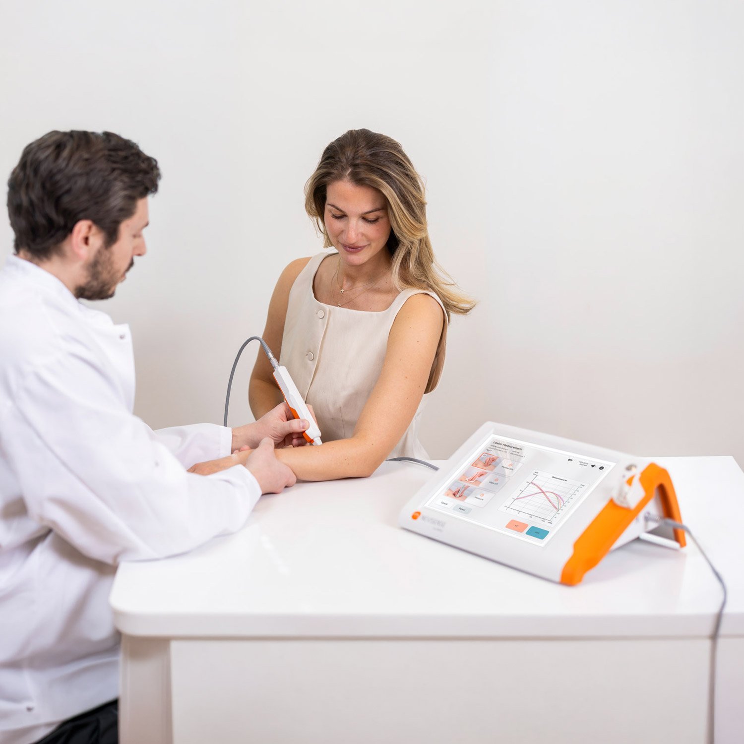
Clinical studies show that Nevisense can increase sensitivity for melanoma detection and provide objective information alongside visual examination and dermoscopy. This additional support can help clinicians make more informed biopsy decisions, particularly when assessing suspicious or ambiguous lesions.
Supporting research and innovation
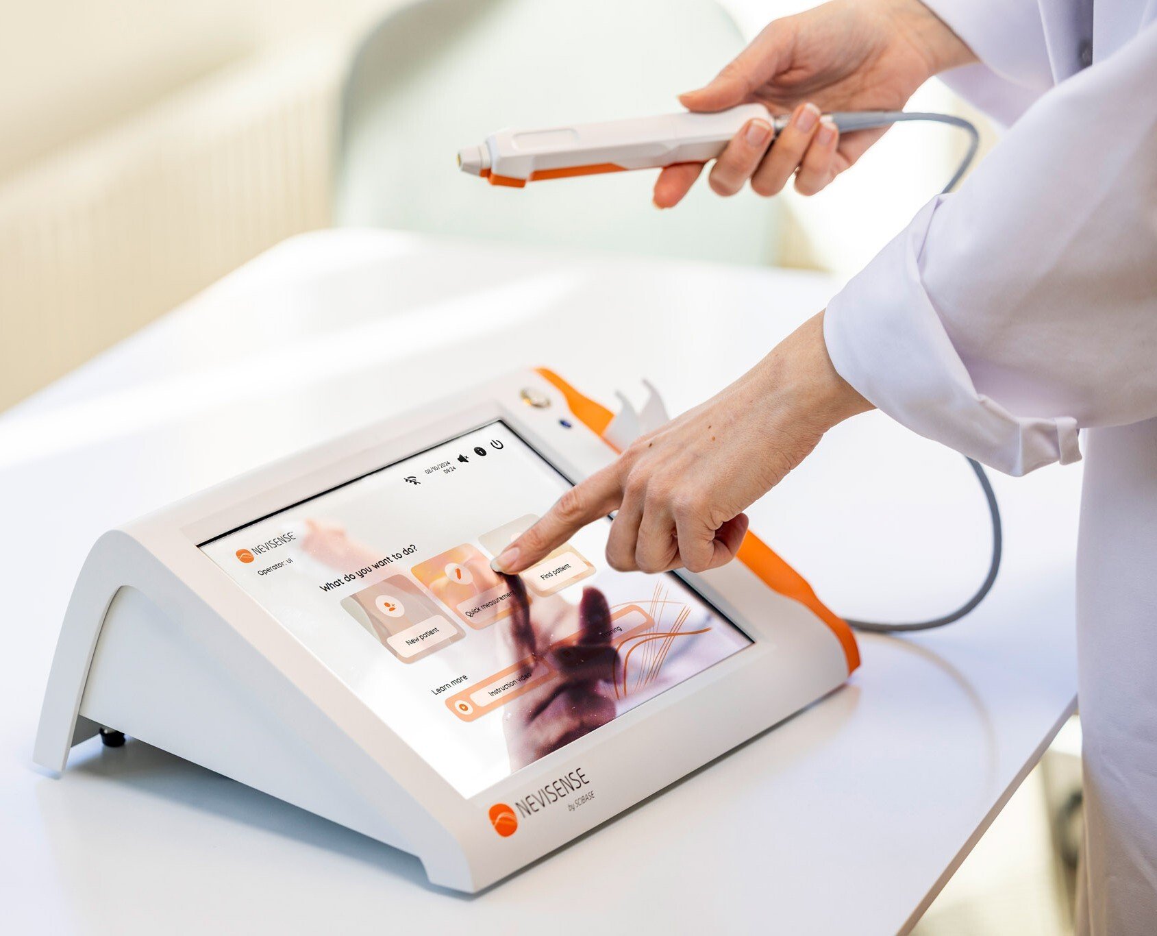
Nevisense is increasingly used in academic studies focused on skin health, barrier function and the effects of dermatological treatments. Research groups value its repeatability, non-invasiveness and quantitative data output.
Used by leading institutions
Hospitals, dermatology clinics and research centers in Germany, the US and other countries have integrated Nevisense into their workflows – citing improved diagnostic decision-making and streamlined patient care.
Trusted by experts. Chosen for results.

Validated in the largest study of its kind
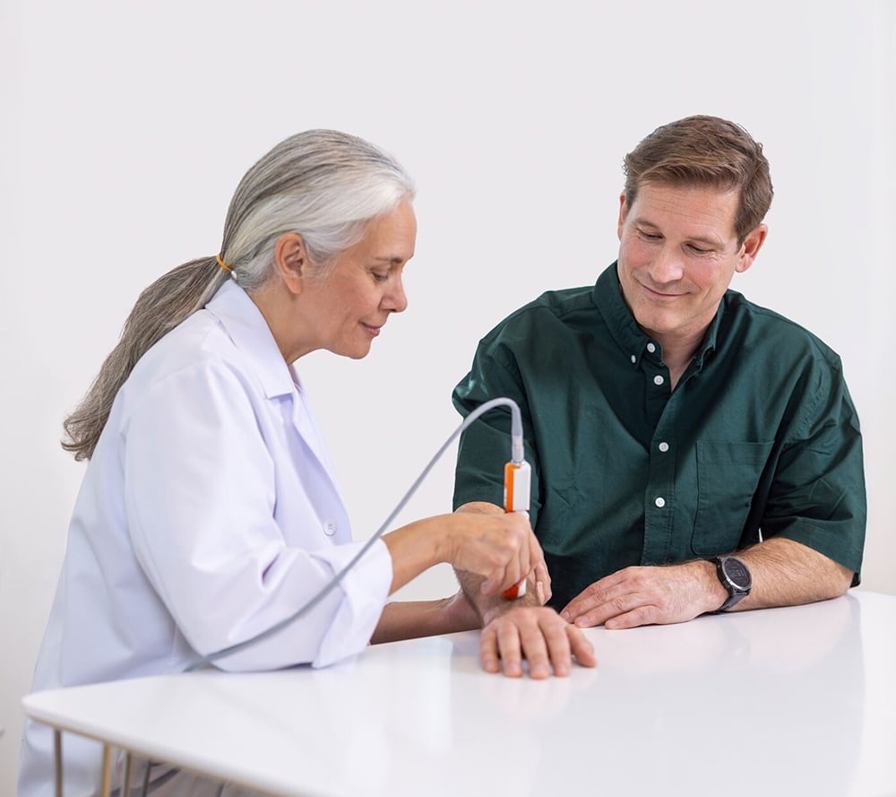
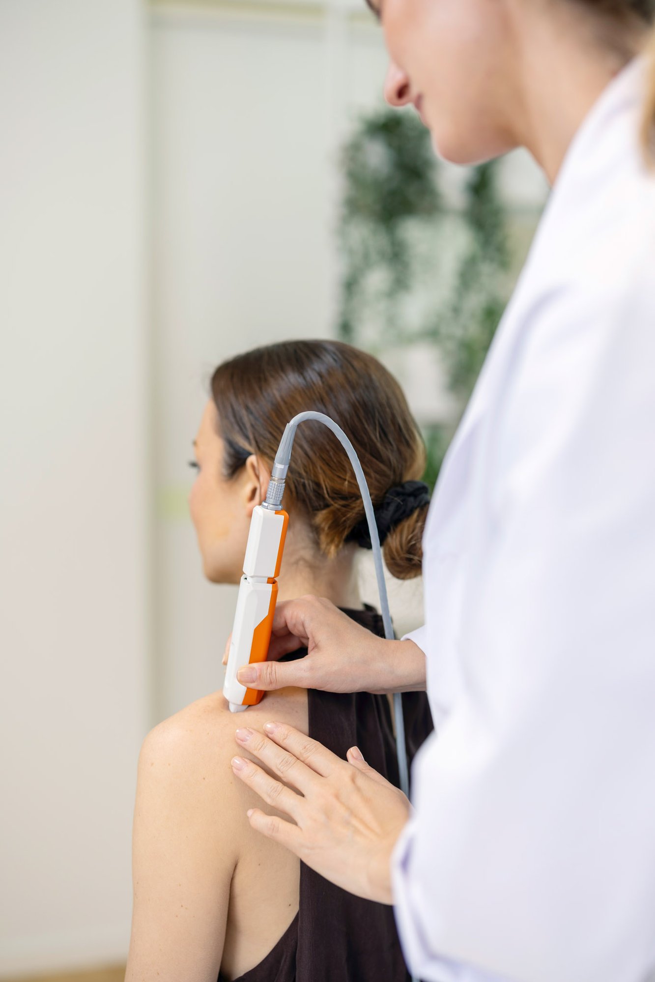
The clinical performance of Nevisense was validated in a pivotal prospective study involving 1,951 patients and 265 melanomas—the largest study of its kind conducted in melanoma detection. The study demonstrated 97% sensitivity and a 99% negative predictive value, providing robust evidence for the use of Nevisense as an adjunct to clinical and dermoscopic evaluation.
Included in guidelines
Nevisense EIS technology is recognized in European and US clinical guidance as a complementary technology that can support the assessment of suspicious skin lesions.
German S1 Guideline
The first German guideline on imaging diagnostics for skin diseases describes EIS as a potentially valuable clinical decision-support tool for assessing melanocytic and keratinocytic lesions.
Read the guideline
EADO Melanoma Guideline
The European melanoma guideline reports a sensitivity of 97.7% and describes EIS as a potentially useful complementary tool in clinical practice.
Read the guideline
US Expert Consensus
A US expert panel concluded that EIS can significantly enhance diagnostic assessment and clinical decision-making for clinically indeterminate pigmented lesions—including melanoma—with SORT Level A evidence.
Read the consensus report
NCCN Guidelines
The NCCN Guidelines identify EIS among the diagnostic-support technologies that may aid in detecting new primary melanoma, particularly in patients with numerous or clinically atypical nevi.
Visit NCCN